Available 24 x 7 x 365
Use our patient portal to easily schedule your exams. Choose a convenient appointment time for you. Now accepting online appointments for:
CT DEXA Mammography MRI Ultrasound X-Ray
Schedule - New Patient Schedule - Established PatientCall Us
Scheduling Phone
(480) 455-1850
Scheduling Hours
Monday–Friday 7:30 am–6:00 pm
Saturday 8:00 am–12:00 pm
Sunday Closed
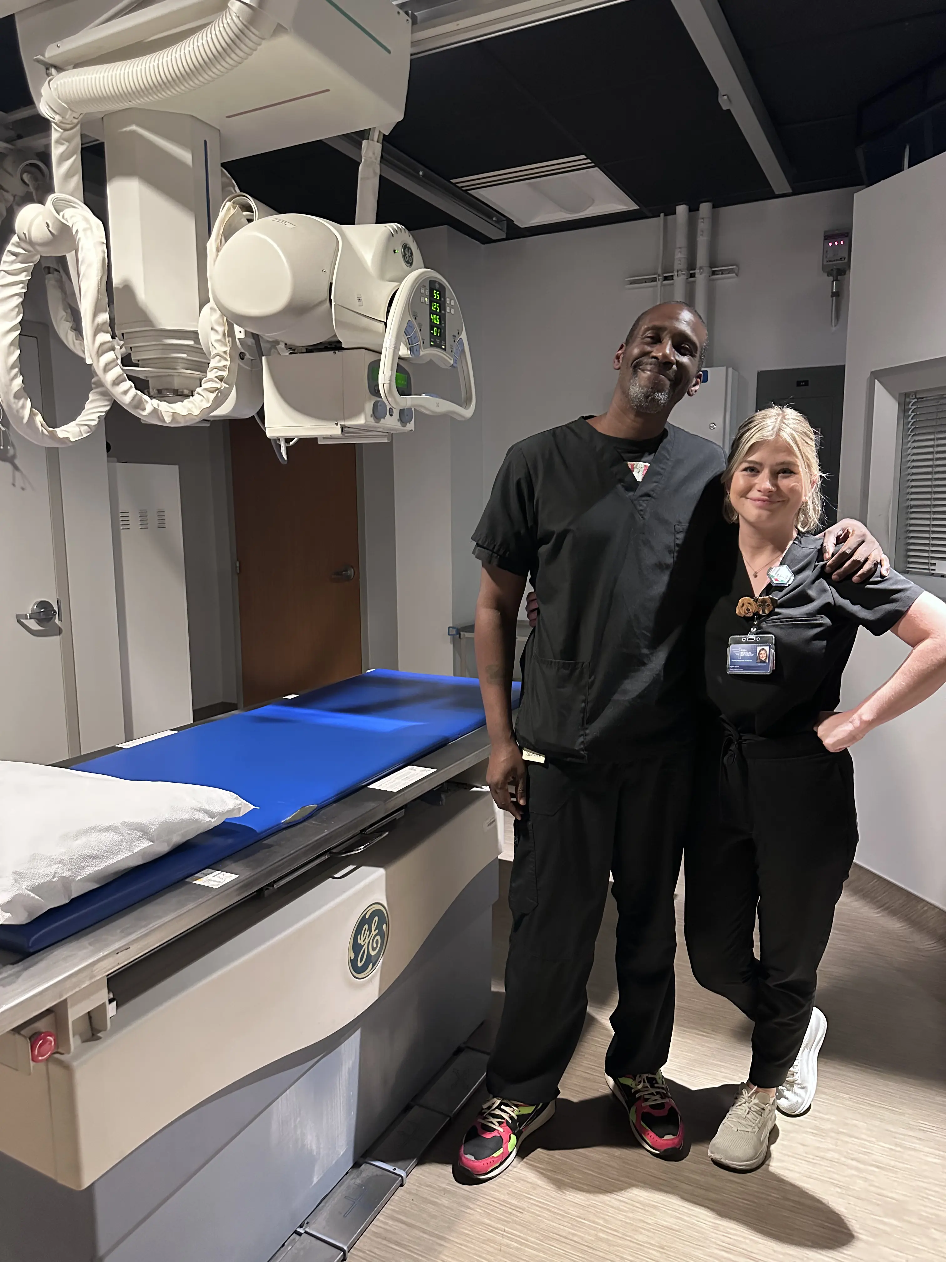
X-Rays
X-Rays can be performed on a walk-in basis.
Visit xrayhours.com for locations, hours, and wait times.
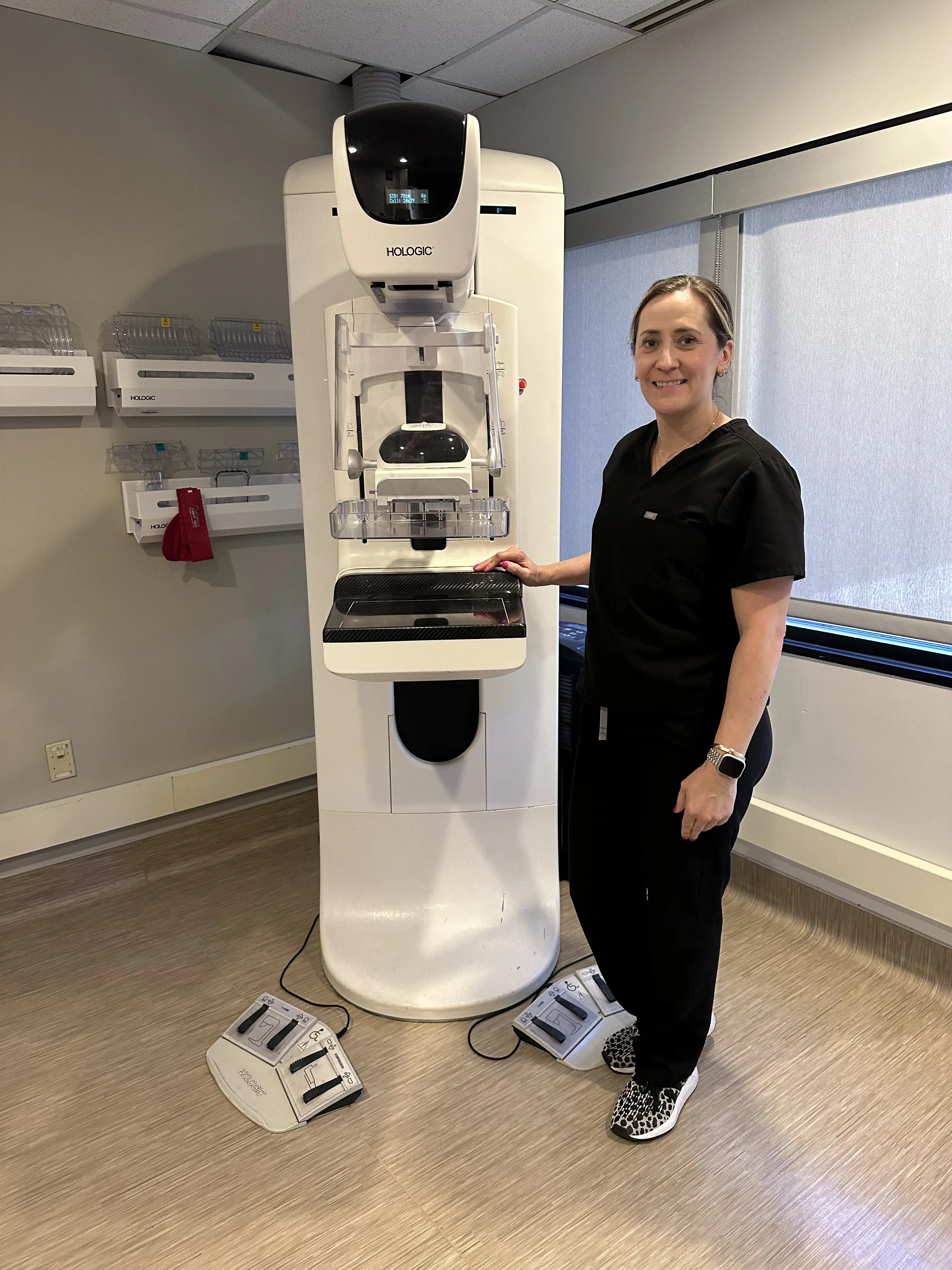
Mammograms
Walk-In 3D Screening Mammograms can be scheduled at
mammohours.com. No doctor order required – just have an active primary care provider or OBGYN.
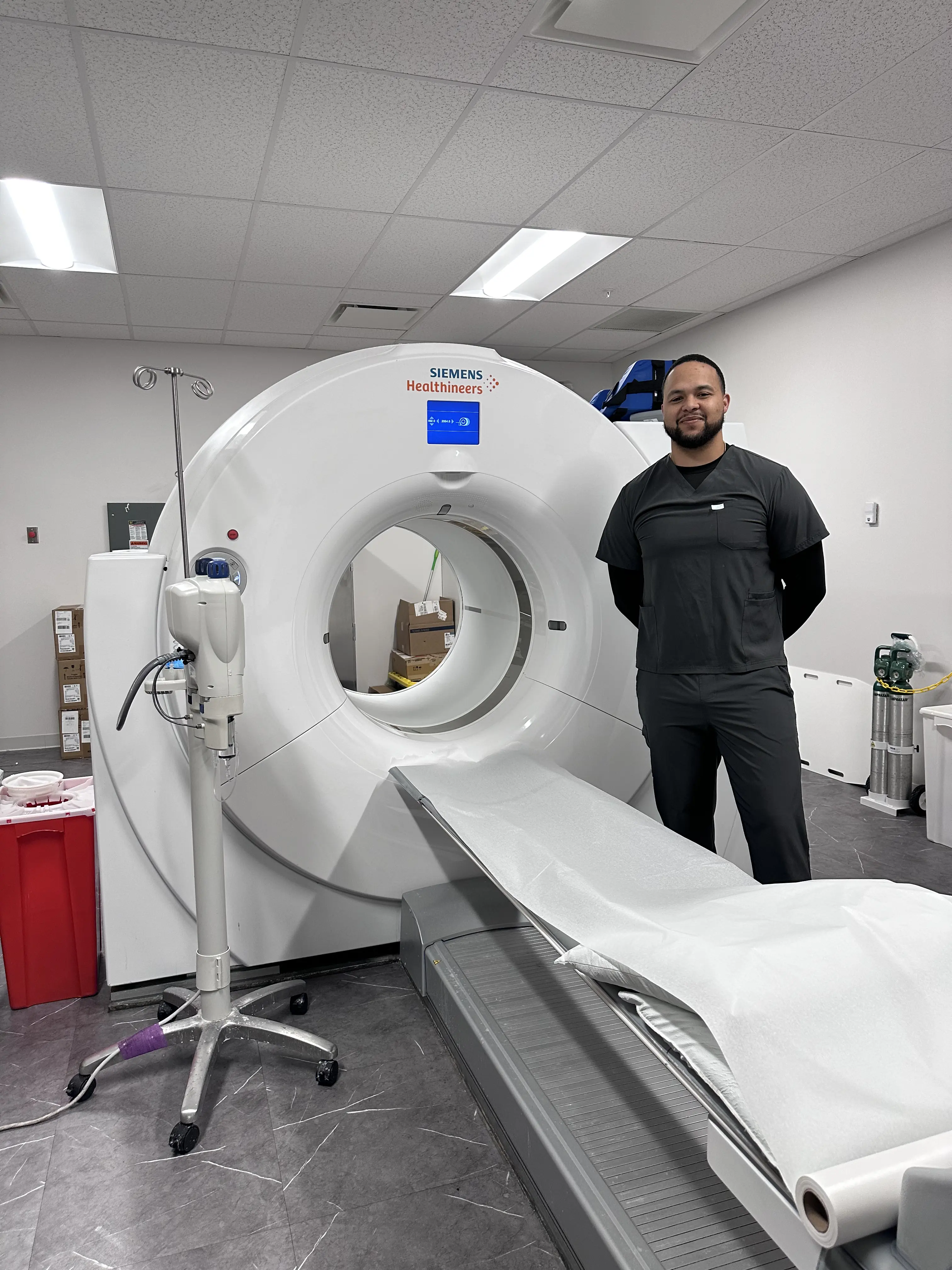
Prostate MRI
A Prostate MRI (magnetic resonance imaging) is a safe, painless test that uses magnetic fields and radio waves to create detailed pictures of the prostate and surrounding soft tissues. Many patients want to know how long a prostate MRI takes. The exam usually takes about 25–30 minutes to complete.
What Does an MRI Show for the Prostate?
During the MRI, you will lie on a table while the scanner takes images of your prostate. The procedure is non-invasive and does not use radiation. The images produced help doctors detect cancer, determine how advanced it may be, and see if it has spread to other areas. If you’re wondering what a prostate MRI shows, it can also reveal other conditions such as benign prostate hyperplasia (BPH) and provide detailed insight into tissue structure and function.
Why a Prostate MRI Is Important
Research shows that performing a prostate MRI before a biopsy in men with high PSA scores can reduce prostate cancer death rates by nearly 20% (The Journal of the American Medical Association). According to the American Cancer Society, 1 in 8 men will be diagnosed with prostate cancer in their lifetime.
While prostate cancer is the second leading cause of cancer death among men in the U.S., survival rates are very high. The 5-year survival rate for prostate cancer in the United States is 99%. Detecting prostate cancer early with prostate MRI increases the odds of successful treatment and outcomes.
Expertise and Accuracy
RadNet, parent company of Arizona Diagnostic Radiology, was one of the first practices in the U.S. to create a dedicated Prostate Program. Our team has performed more prostate MRI exams than nearly any other group, ensuring accuracy that meets or exceeds top academic centers. If you are searching for a prostate MRI near you, our Arizona locations provide accessible, state-of-the-art imaging.
Patients also often ask about prostate MRI costs in Phoenix, AZ, and our staff provides clear, upfront guidance on pricing and insurance coverage.
A prostate MRI is a valuable tool for early detection and careful monitoring, giving men and their doctors the information needed to make informed decisions about treatment and follow-up care.