Available 24 x 7 x 365
Use our patient portal to easily schedule your exams. Choose a convenient appointment time for you. Now accepting online appointments for:
CT DEXA Mammography MRI Ultrasound X-Ray
Schedule - New Patient Schedule - Established PatientCall Us
Scheduling Phone
(480) 455-1850
Scheduling Hours
Monday–Friday 7:30 am–6:00 pm
Saturday 8:00 am–12:00 pm
Sunday Closed
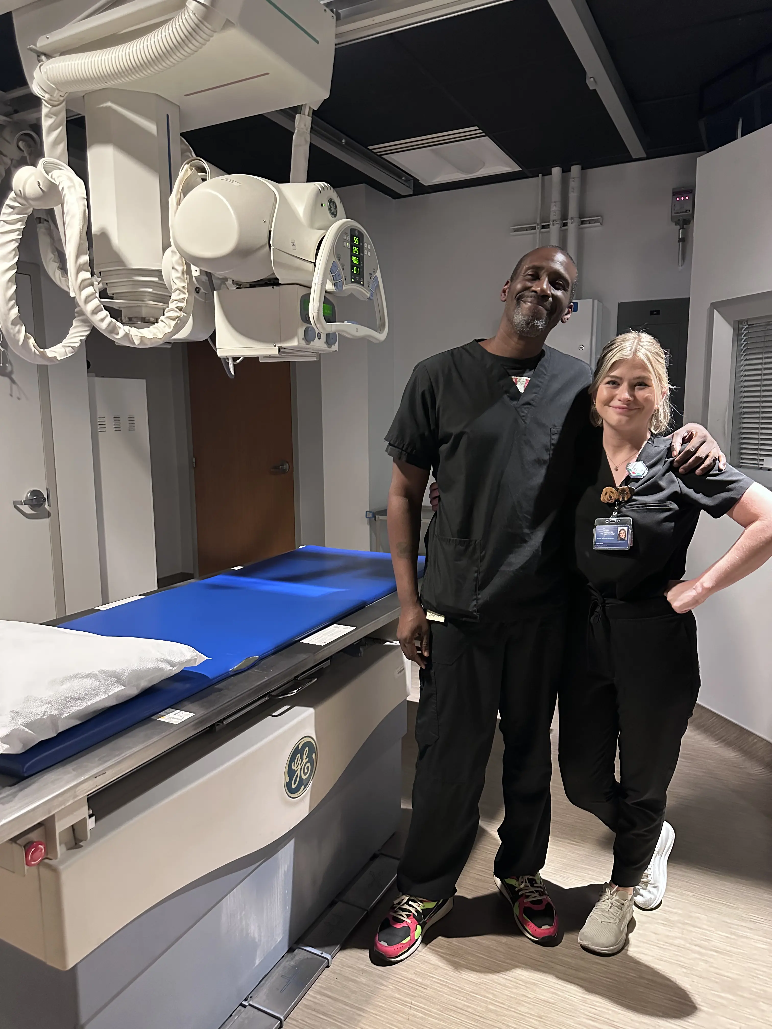
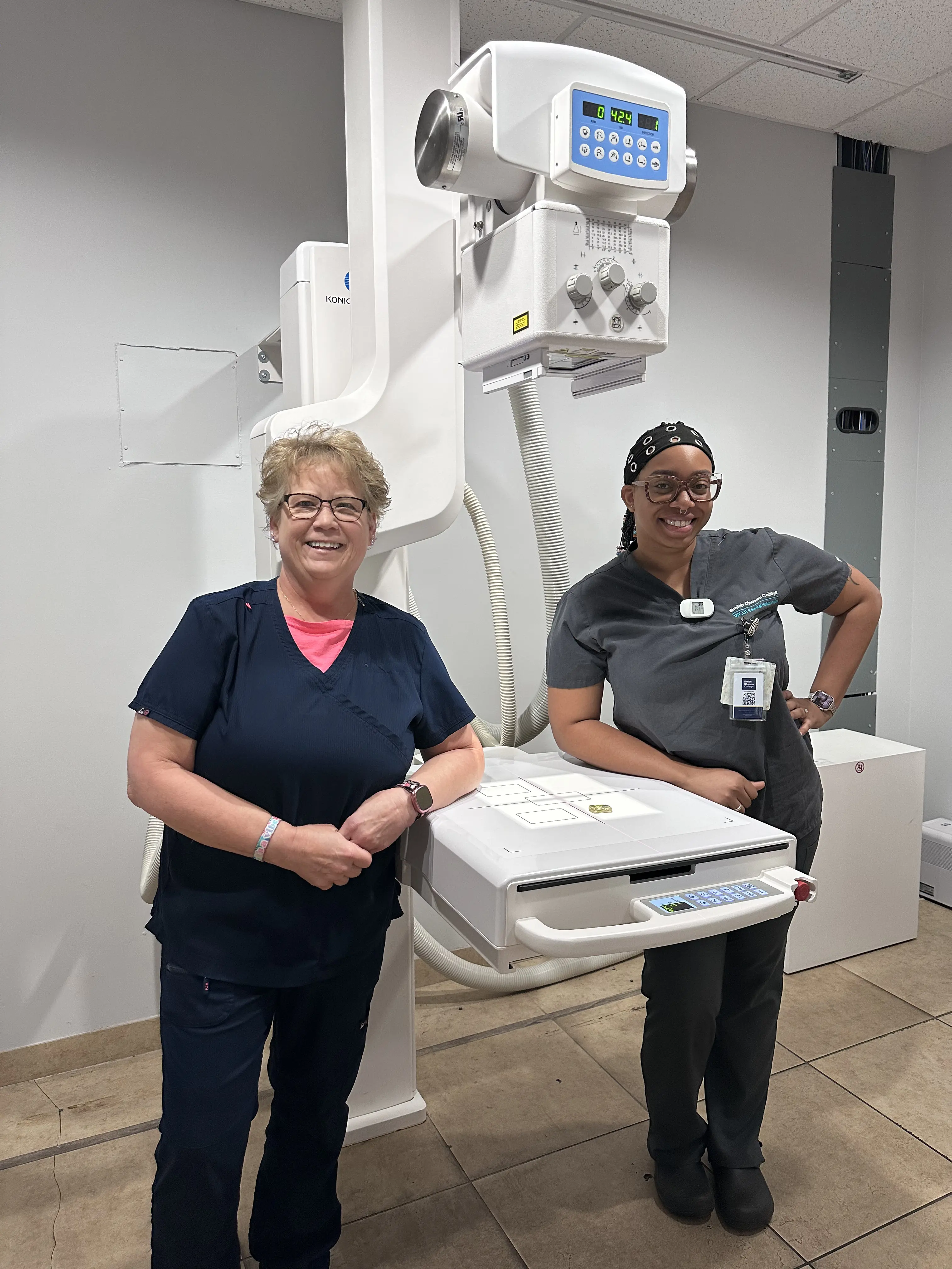
X-Rays
X-Rays can be performed on a walk-in basis.
Visit xrayhours.com for locations, hours, and wait times.
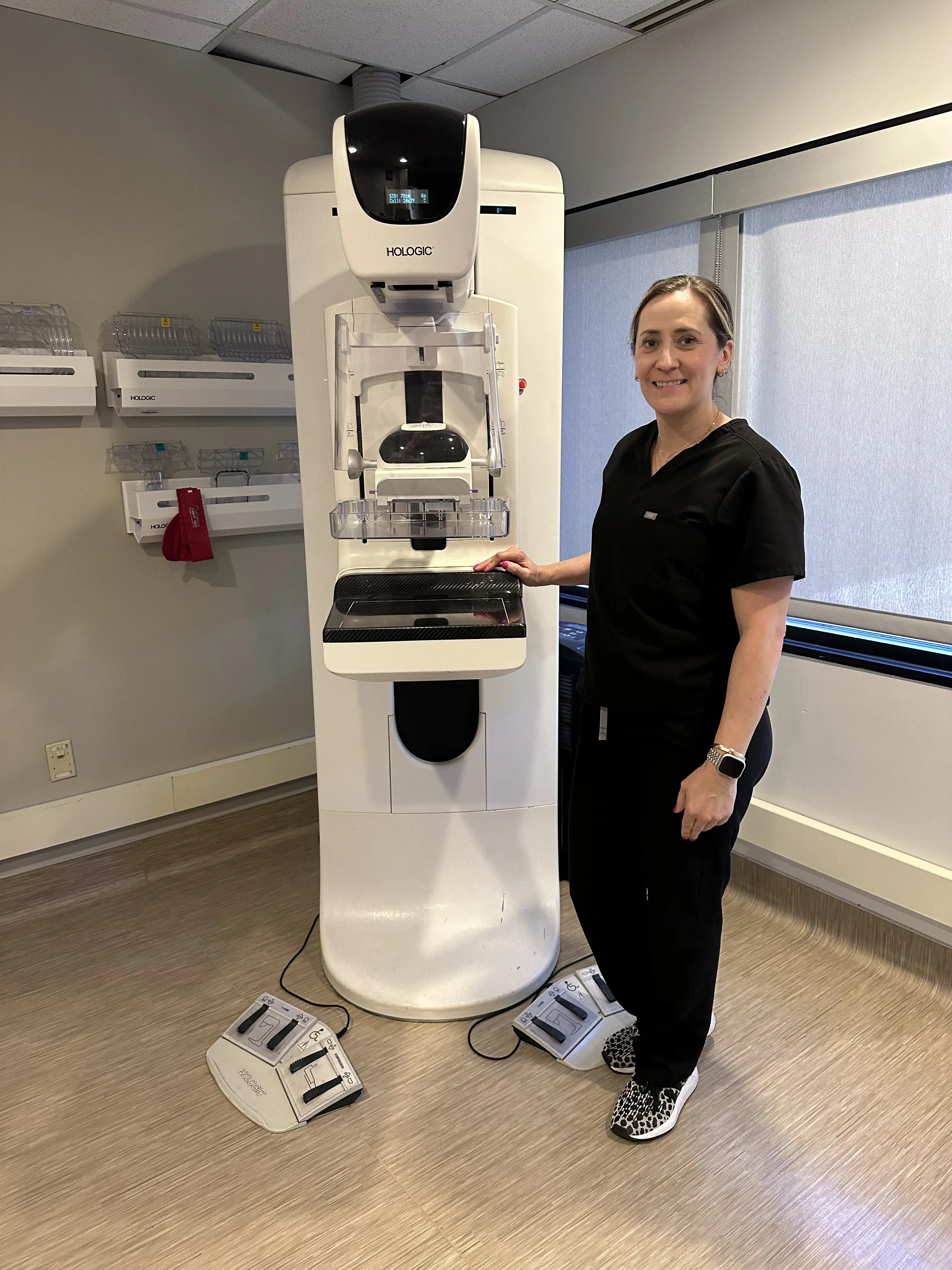
Mammograms
Walk-In 3D Screening Mammograms can be scheduled at
mammohours.com. No doctor order required – just have an active primary care provider or OBGYN.
Nuclear Medicine
Nuclear medicine is a type of imaging that uses a small amount of radioactive material, called a radiotracer, to take detailed pictures inside your body. After the tracer is given, a PET/MRI or PET/CT scan detects the radiation it gives off and creates computer images. These images are then reviewed by our fellowship-trained radiologists to help diagnose or monitor different health conditions.
What is Nuclear Medicine
Nuclear Medicine uses a radiotracer that is usually injected into a vein in your arm. Once inside your body, it travels to the specific area your doctor wants to study. For example, it may collect in a tumor, an inflamed area, or a certain organ. Tracers can also attach to proteins to help show how your body is working on a cellular level.
Unlike a regular X-ray, which mainly shows bones, nuclear medicine makes it easier to see organs, tissues, and blood flow. The way your body absorbs the tracer helps doctors understand not only what your organs look like, but also how well they are functioning.
Radiotracers are not dyes or medications, and they usually cause no side effects. The amount of radiation you receive is very small, about the same as or less than many standard imaging tests. The tracer naturally decays to a non-radioactive state shortly after your exam and is then flushed out of your system naturally. Drinking plenty of fluids after a nuclear imaging scan helps speed up the removal process.
What Nuclear Medicine Can Detect
Nuclear medicine is commonly used to diagnose or monitor:
- Blood disorders
- Thyroid problems
- Heart disease
- Gallbladder disease
- Lung conditions
- Bone issues, such as fractures or infections
- Kidney disease, including blockages or scars
- Cancer
It can also be used to guide treatments or check how well a treatment is working. Nuclear medicine provides doctors with both images and functional information, giving a more complete view of your health.