Available 24 x 7 x 365
Use our patient portal to easily schedule your exams. Choose a convenient appointment time for you. Now accepting online appointments for:
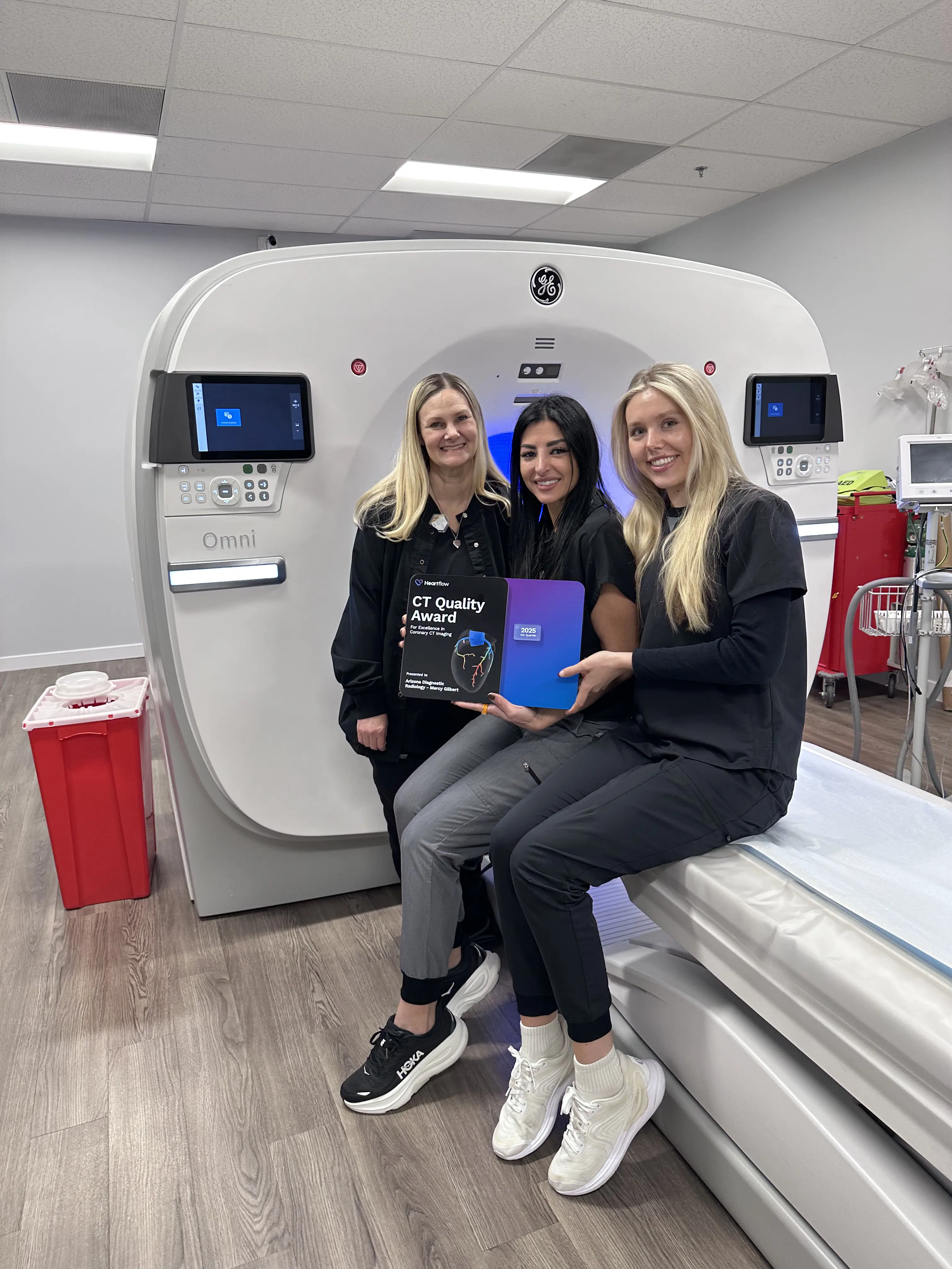
CT DEXA Mammography MRI Ultrasound X-Ray
Schedule - New Patient Schedule - Established PatientCall Us
Scheduling Phone
(480) 455-1850
Scheduling Hours
Monday–Friday 7:30 am–6:00 pm
Saturday 8:00 am–12:00 pm
Sunday Closed
X-Rays
X-Rays can be performed on a walk-in basis.
Visit xrayhours.com for locations, hours, and wait times.
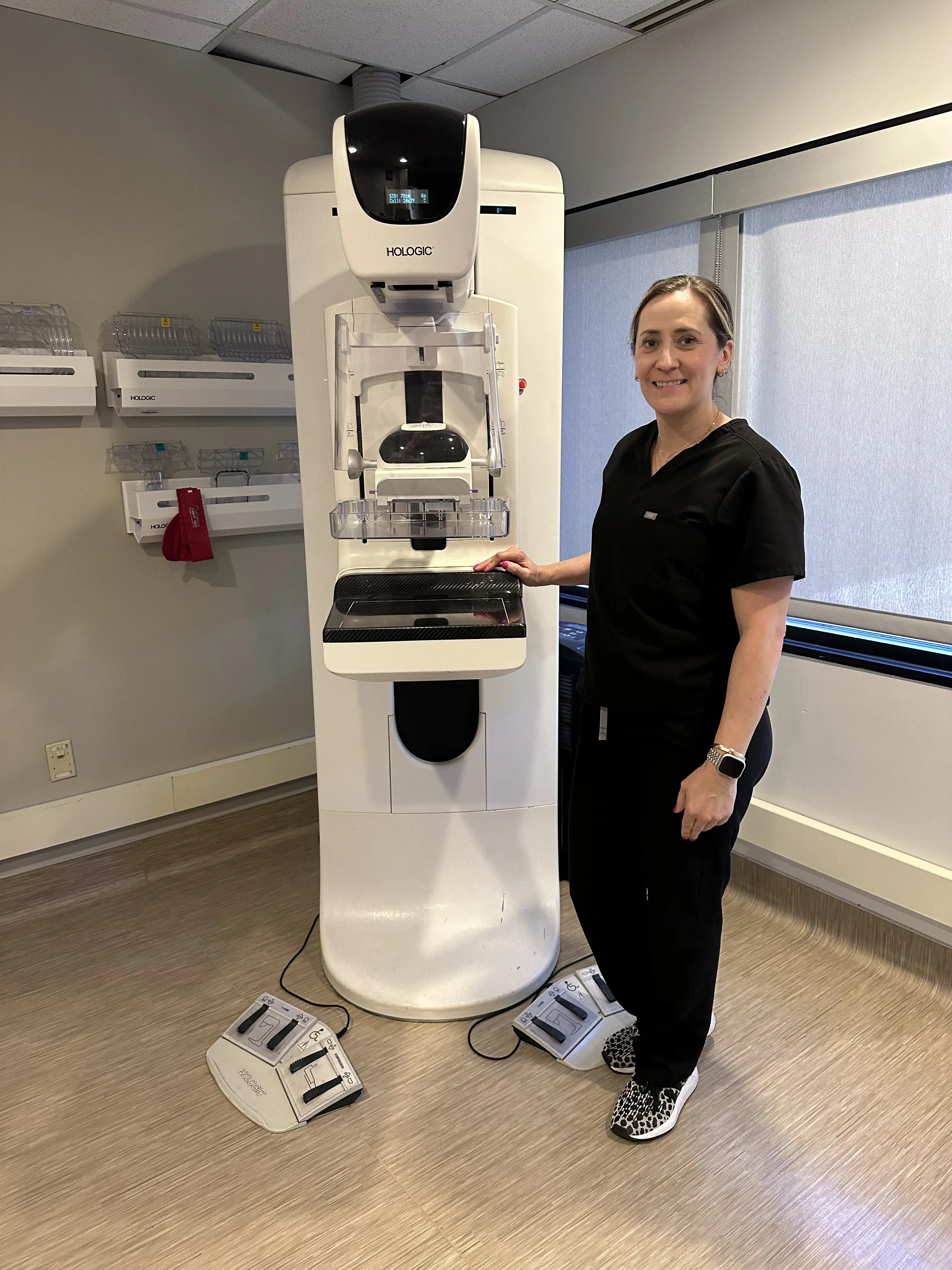
Mammograms
Walk-In 3D Screening Mammograms can be scheduled at
mammohours.com. No doctor order required – just have an active primary care provider or OBGYN.
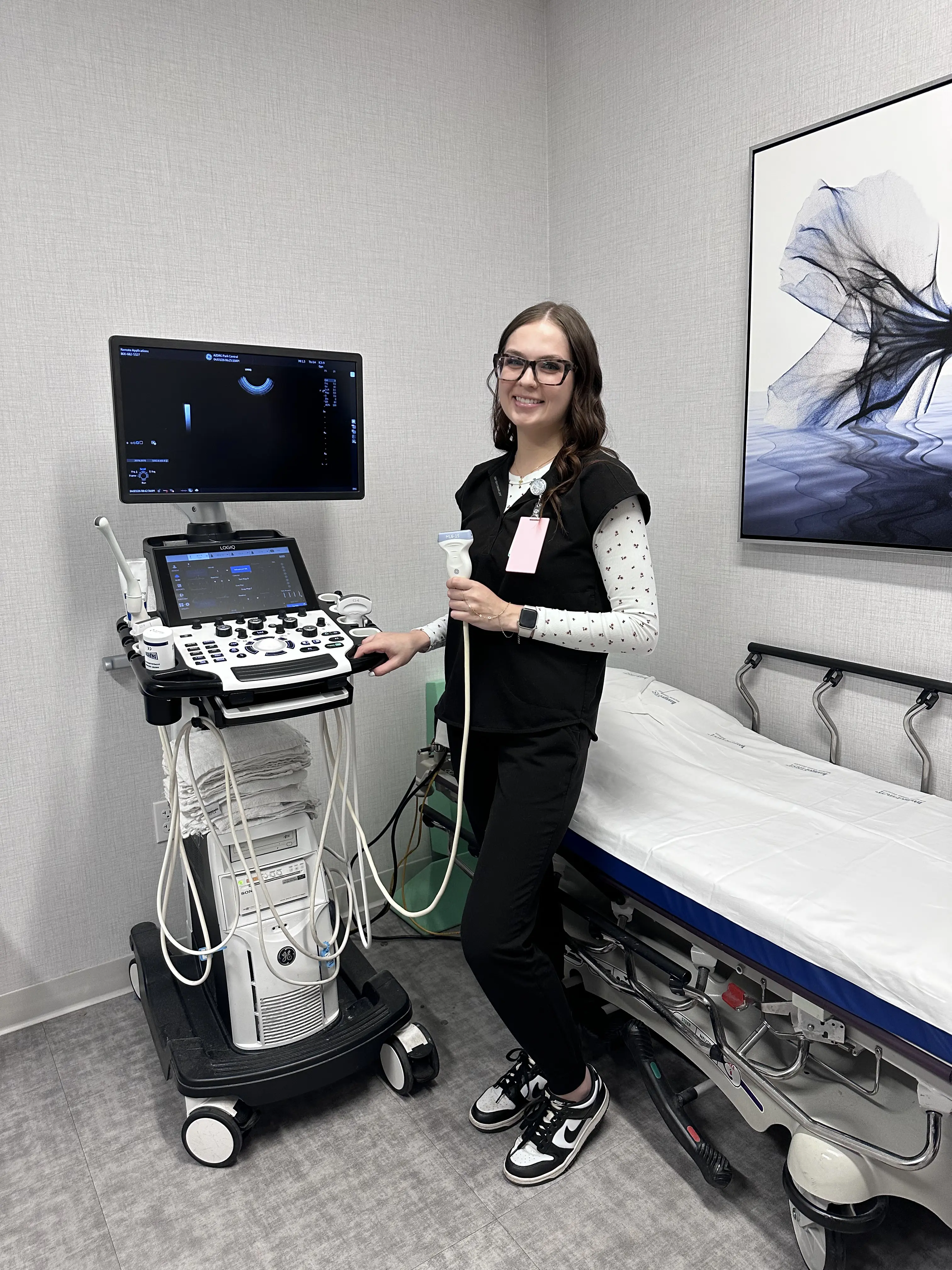
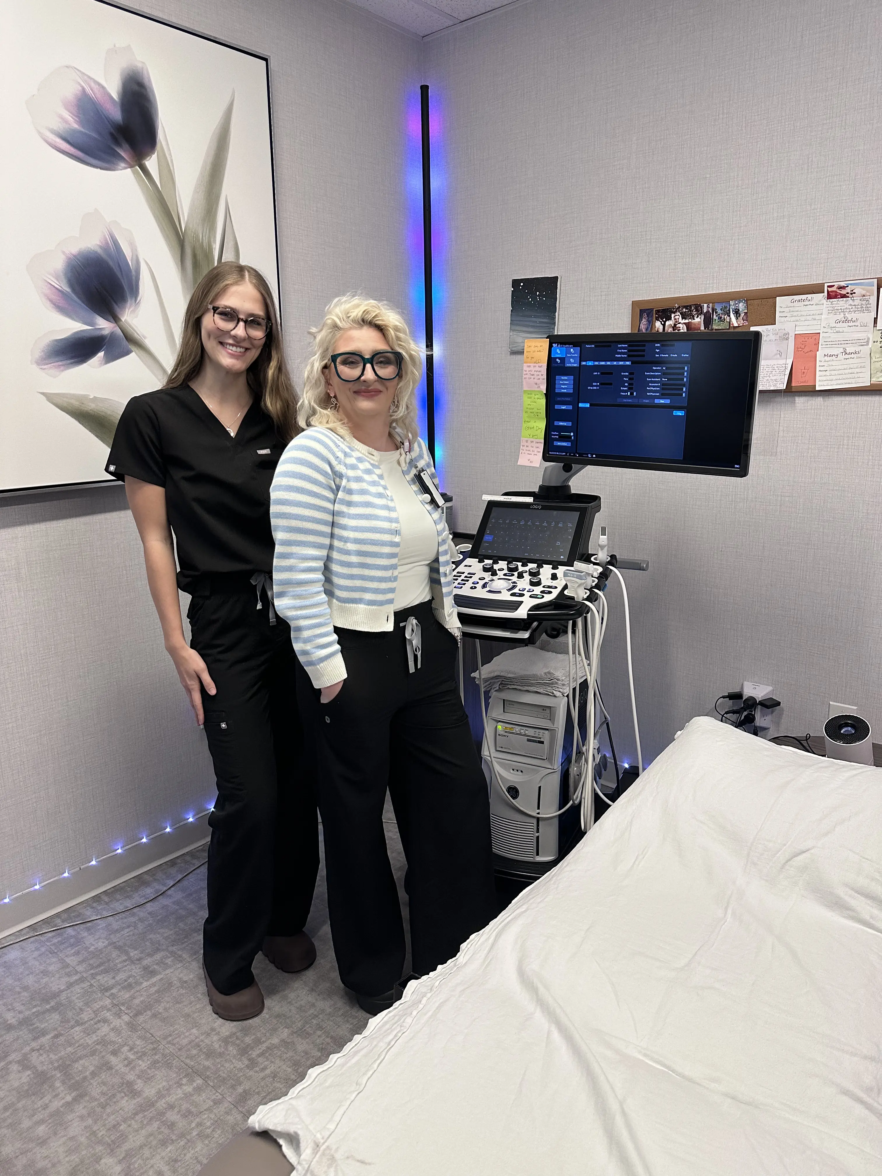
Phoenix Ultrasound
An ultrasound, also called sonography, is a safe and painless test that uses sound waves to make pictures of the inside of your body.
These pictures are called sonograms. These detailed pictures can help your doctor diagnose and monitor many conditions. Unlike X-rays or CT scans, ultrasound does not use radiation. Ultrasound (Phoenix) creates real-time images or video of organs, tissues, and blood vessels. This helps doctors see what is happening inside the body right away.
Ultrasound costs can differ depending on what area is being scanned, if contrast is used, and your insurance plan. Our team can help you understand pricing before your appointment so there are no surprises.
How Ultrasound Works
During the test, a technologist (also known as a sonographer) uses a small device called a transducer or probe. A thin layer of clear gel is placed on your skin so the sound waves can move easily into your body.
The probe sends high-frequency sound waves into your body. These sound waves bounce back when they hit organs and tissues. A computer then changes the echoes into moving images that appear on a nearby screen.
Areas Ultrasound Can Check
Although many people think of ultrasound as tool to monitor a fetus during pregnancy, it is used for many other areas of the body, including:
- Abdomen/Stomach
- Kidneys (renal area)
- Breasts
- Pelvic area
- Thyroid
- Transrectal exams
- Transvaginal exams
Common Uses of Ultrasound
Ultrasound has many uses beyond pregnancy. Doctors often use it to:
- Find the cause of pain, swelling, or infection
- Detect gallstones or tumors
- Check arteries and veins for narrowing, blockages, or clots
- Examine organs like the liver, kidneys, and bladder
If you’re researching ultrasound versus other imaging services, our technologists can explain which is best for your needs and how each test helps your doctor get accurate results.
What to Expect During Your Exam
You will lie on a comfortable exam table. The sonographer will put gel on the skin over the area being checked. The transducer is moved gently over your skin to take images.
How long does an ultrasound take?
Most exams are very quick, usually lasting less than 30 minutes. Once it’s done, you can get dressed and leave right away, with no recovery time needed.
If you have more questions, our staff is happy to provide details and help you prepare for your visit.
How Much Does an Ultrasound Cost in Phoenix?
If you’re wondering how much an ultrasound costs in Phoenix, the price can vary depending on several factors such as the type of ultrasound exam, the area of the body being scanned, and your individual insurance coverage, co-pay, or deductible.
That’s why many patients choose outpatient imaging centers like Arizona Diagnostic Radiology, where you can receive the same high-quality diagnostic ultrasound at a much lower cost than hospitals. According to the National Institute for Health Care Reform, community-based imaging centers may charge up to 50% less than hospital outpatient departments for identical exams.
Choosing an outpatient facility not only helps you save money but also ensures fast scheduling, comfortable service, and expert results you can trust.
If you’re looking for an ultrasound in Phoenix, Arizona Diagnostic Radiology offers affordable imaging, combining advanced technology, compassionate care, and clear, accurate answers.