Available 24 x 7 x 365
Use our patient portal to easily schedule your exams. Choose a convenient appointment time for you. Now accepting online appointments for:
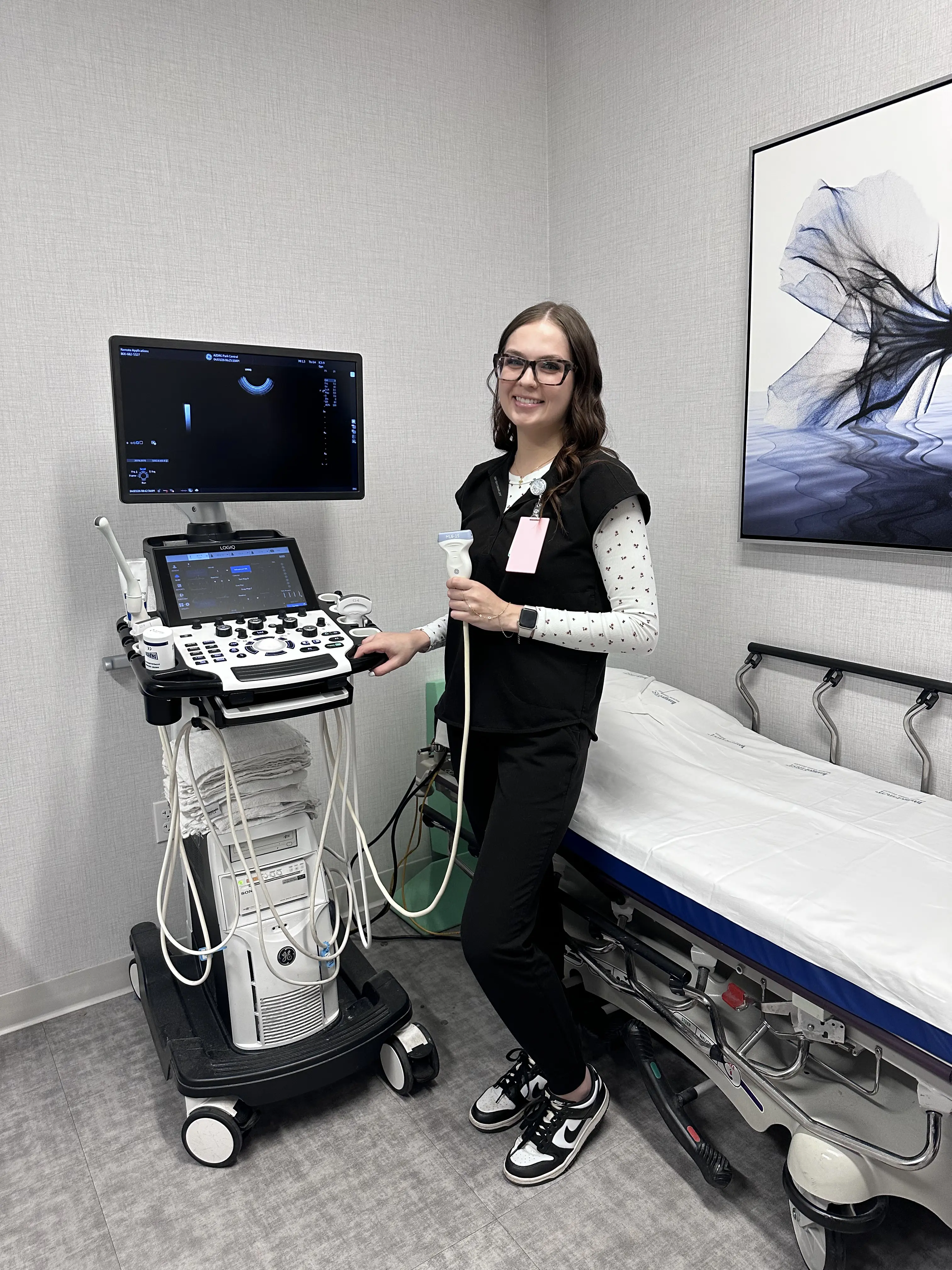
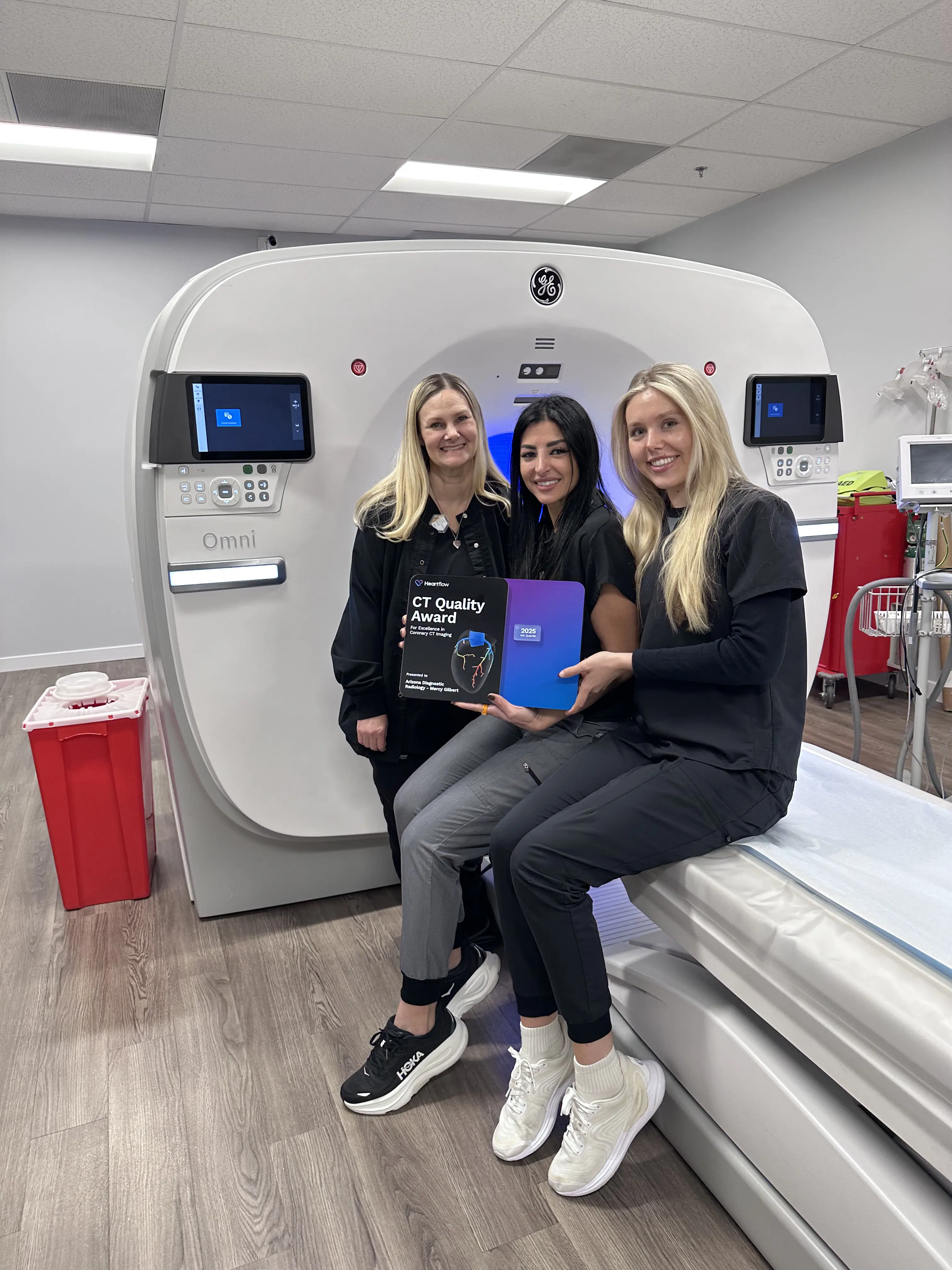
CT DEXA Mammography MRI Ultrasound X-Ray
Schedule - New Patient Schedule - Established PatientCall Us
Scheduling Phone
(480) 455-1850
Scheduling Hours
Monday–Friday 7:30 am–6:00 pm
Saturday 8:00 am–12:00 pm
Sunday Closed
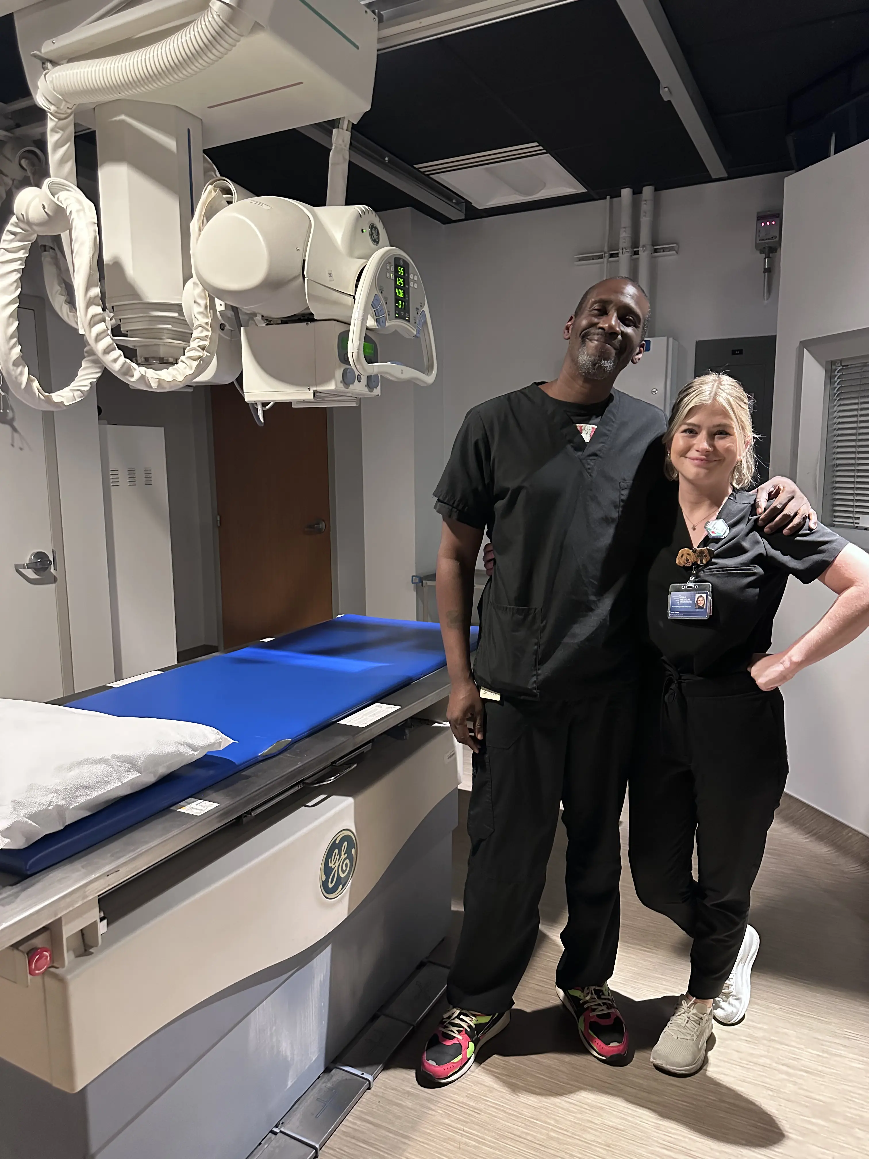
X-Rays
X-Rays can be performed on a walk-in basis.
Visit xrayhours.com for locations, hours, and wait times.
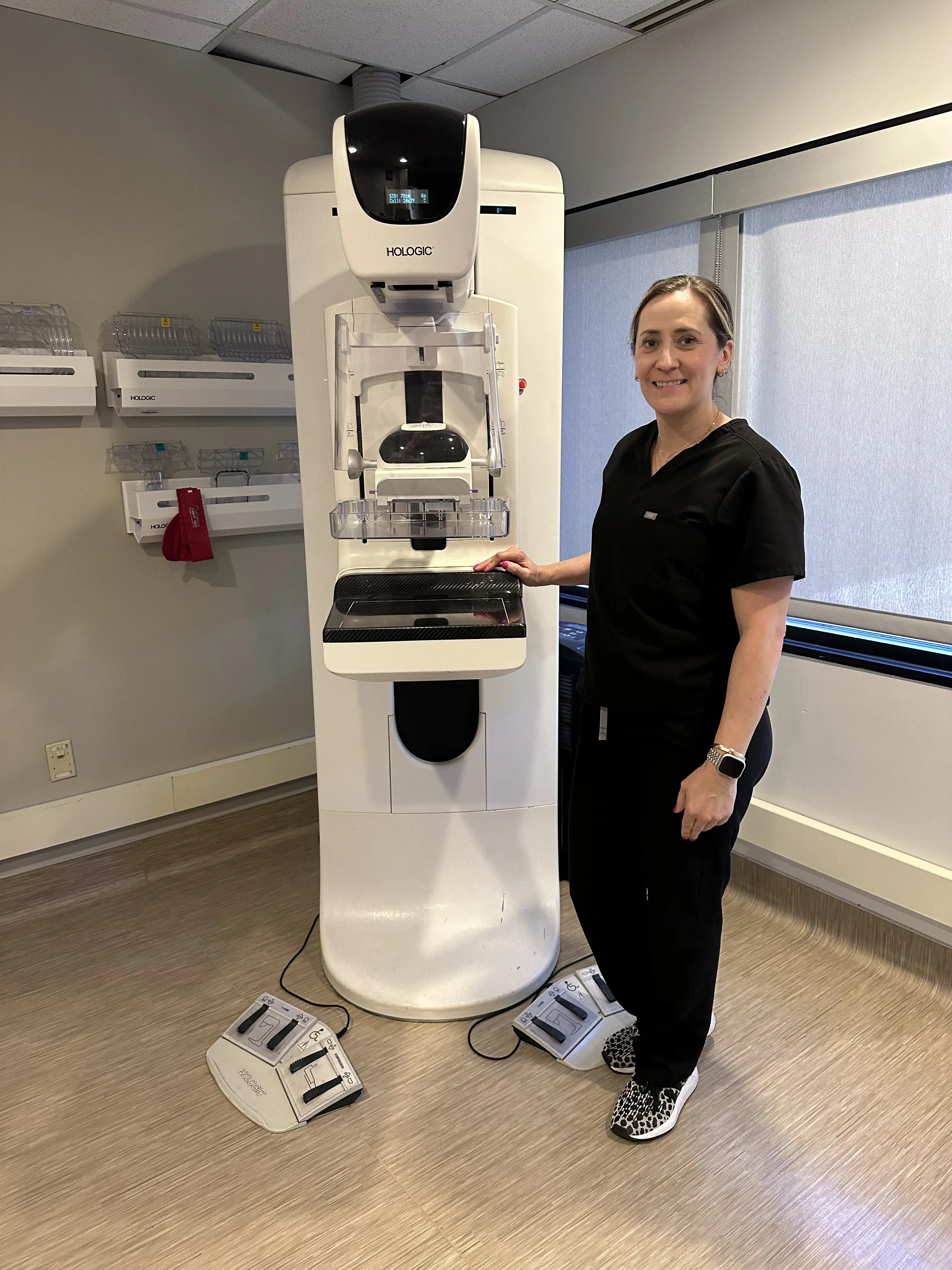
Mammograms
Walk-In 3D Screening Mammograms can be scheduled at
mammohours.com. No doctor order required – just have an active primary care provider or OBGYN.
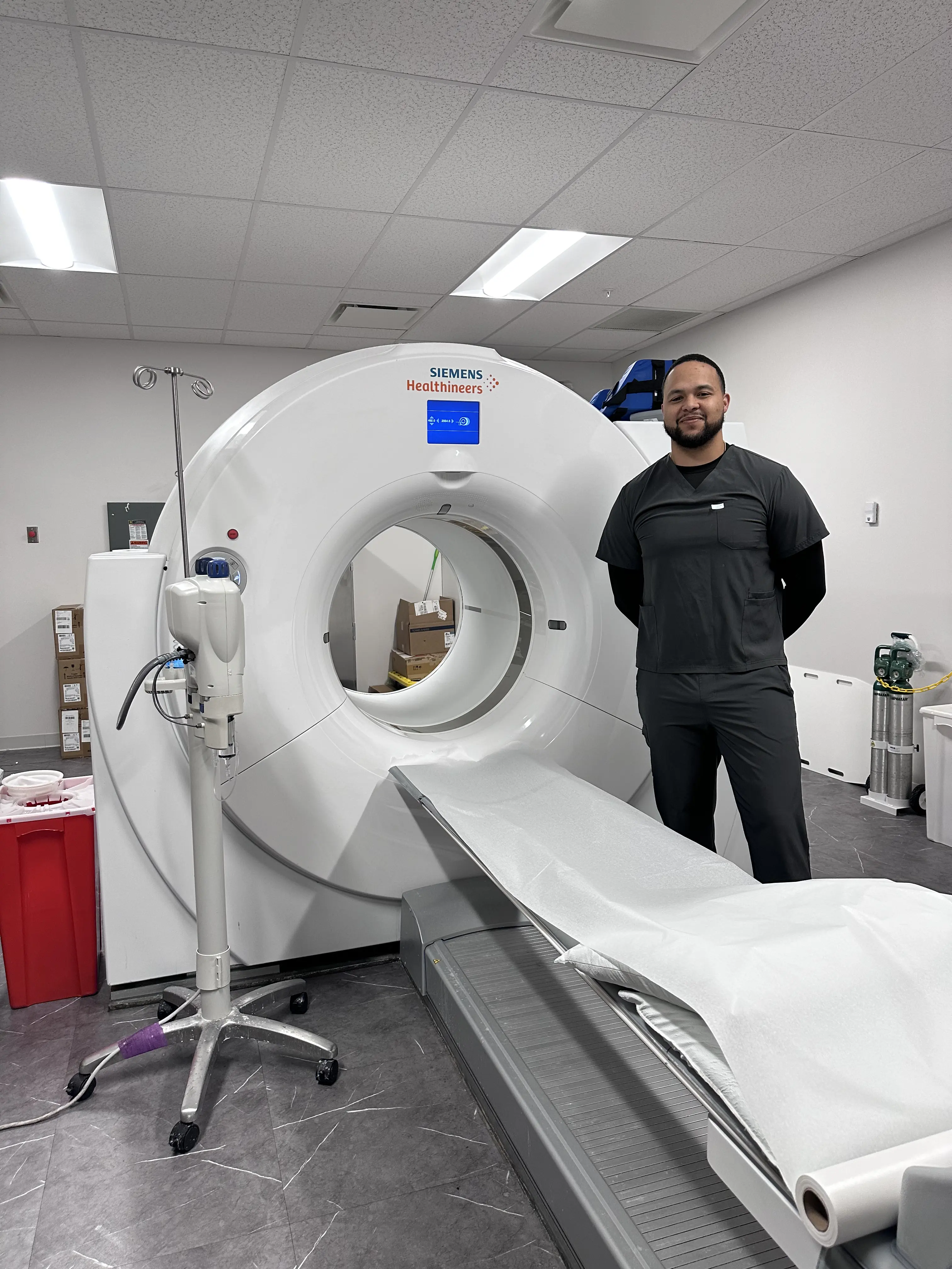
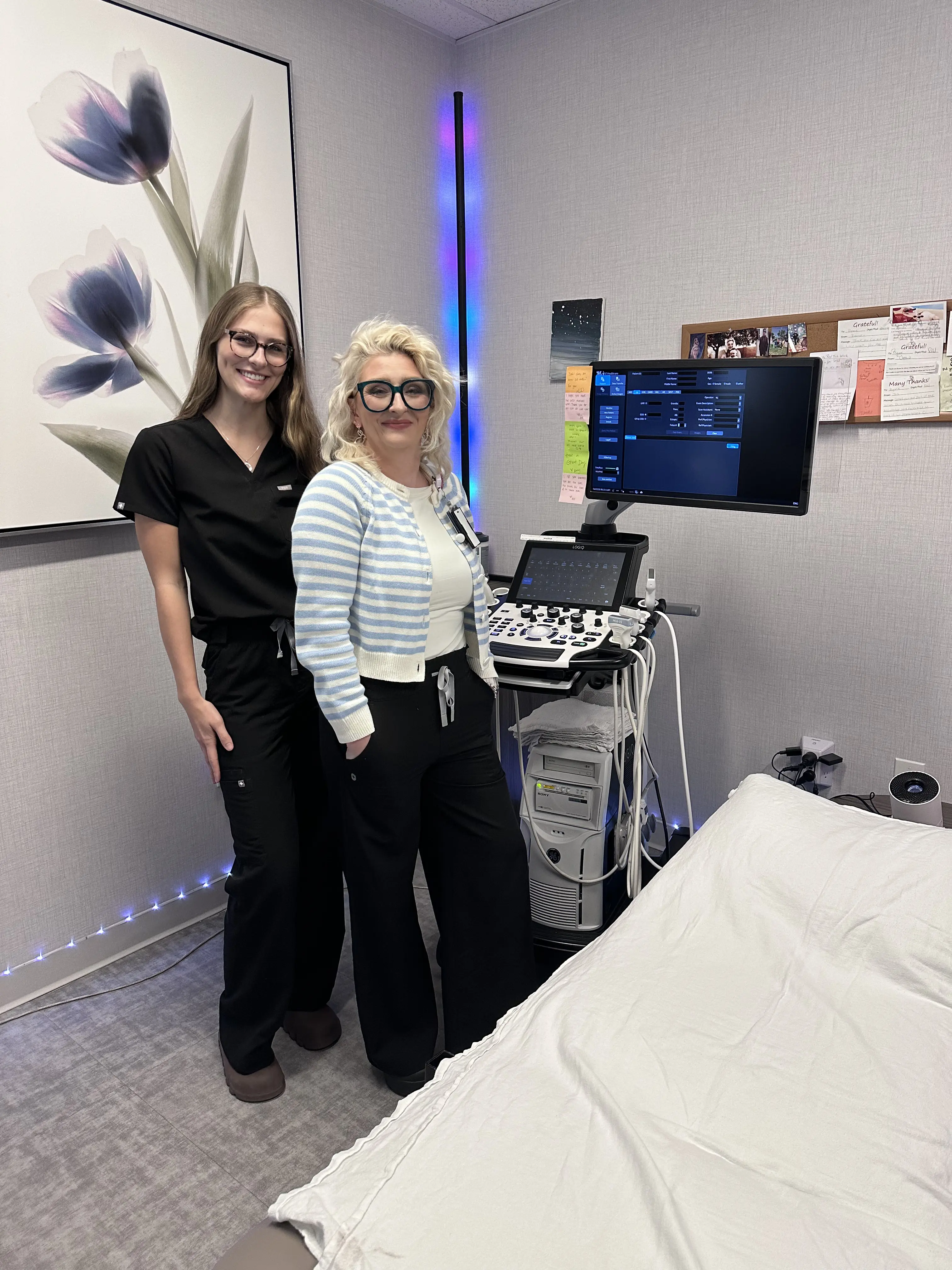
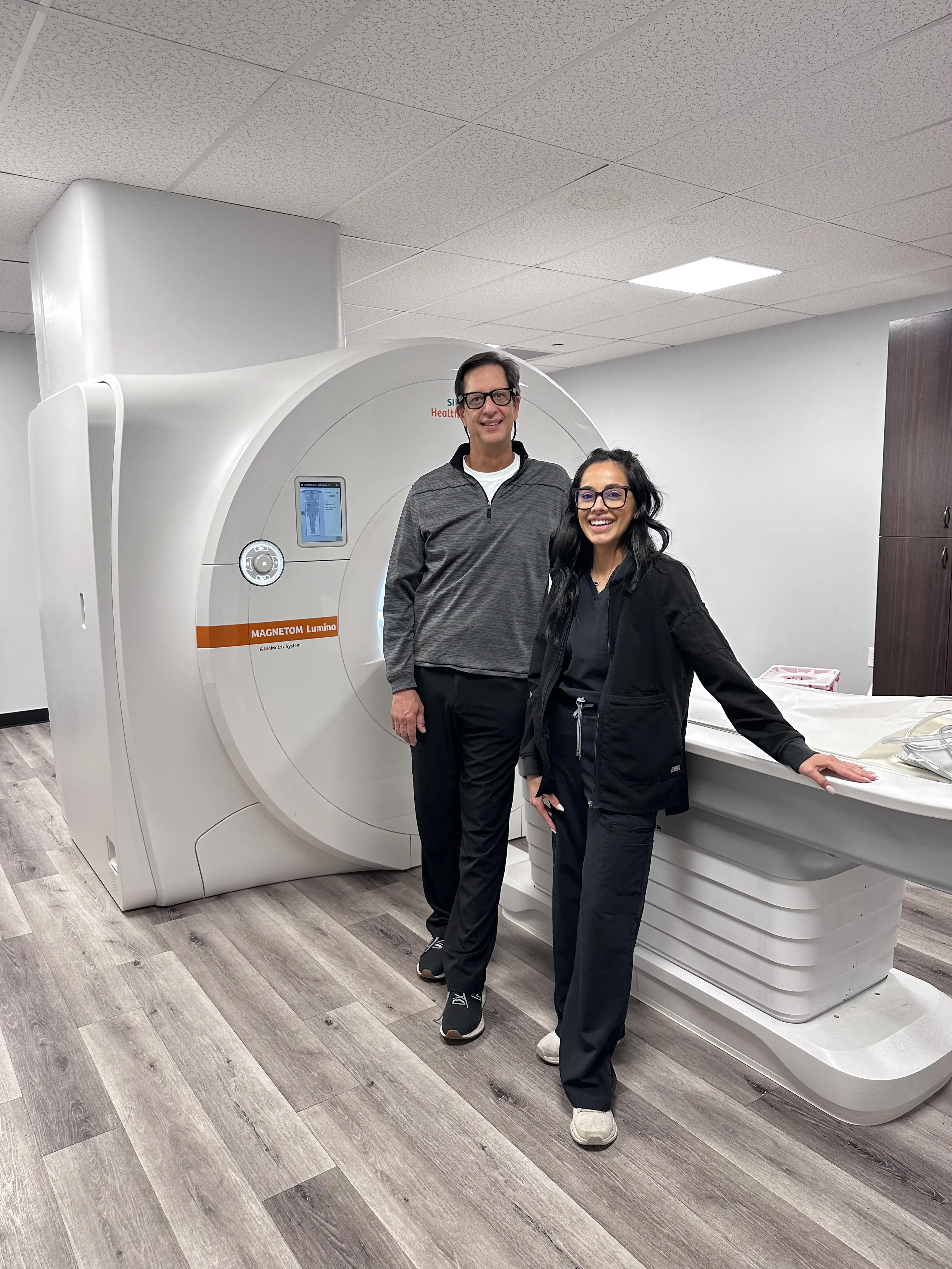
Breast MRI
A Breast MRI (magnetic resonance imaging) is a type of imaging test that uses a large magnet, radio waves, and a computer to create detailed pictures of breast tissue. Unlike a mammogram, a breast MRI does not use X-rays, so it contains no radiation. It is often recommended in addition to mammograms for patients with dense breast tissue who may need more advanced imaging.
How Breast MRI Works
During a breast MRI, you will lie on a table while the scanner takes images of your breast tissue. Some exams may use a contrast dye injected into your vein. The dye helps the radiologist see normal and abnormal areas more clearly. Not every breast MRI requires contrast; it depends on why the test is being done.
What Does an MRI of the Breast Show?
Breast MRI is often used along with a mammogram, not as a replacement. It may be recommended for people who need more detailed imaging, including:
- High-Risk Breast Program – If you have a strong family history of breast cancer, carry genes that increase risk, or have dense breast tissue
- Evaluating new breast cancer – To see the size of a tumor or extent of cancer growth
- Checking breast implants – To look for possible ruptures in silicone implants
Many patients wonder about breast MRI screening guidelines, as these exams are often suggested for high-risk patients or those with dense breast tissue. If you are unsure whether you qualify for screening, your doctor can review your personal and family history to determine the best approach.
Breast MRI can detect cancers that mammograms or ultrasounds may miss, especially in people with dense breast tissue. It gives your doctor detailed information to make the best treatment and screening decisions for you.
What to Expect
Breast MRI is safe, painless, and noninvasive. Most people can return to normal activities immediately after the exam. Your care team will explain whether a Breast MRI with contrast dye will be used and answer any questions about the procedure.
A breast MRI vs. mammogram discussion is common. While mammograms remain the gold standard for screening, breast MRI can be a vital adjunct screening tool, providing more detailed images, especially for high-risk individuals. A breast MRI can help doctors find cancer early, evaluate breast implants, and plan treatment with confidence.
How Much Does a Breast MRI Cost in Arizona?
If you’re wondering how much a breast MRI costs in Arizona, the price can vary depending on whether contrast is used, the type of MRI, and your insurance coverage, co-pay, or deductible. Hospital-based facilities often charge significantly more for the same procedure, sometimes up to twice as much.
Many patients choose outpatient imaging centers like Arizona Diagnostic Radiology to receive the same high-quality breast MRI exams at a fraction of the cost. According to the National Institute for Health Care Reform, community-based imaging centers may charge up to 50% less than hospital outpatient departments for identical procedures (source).
Outpatient facilities not only provide substantial savings, but also faster scheduling, personalized care, and a comfortable experience without compromising accuracy.
Arizona Diagnostic Radiology offers affordable breast MRI imaging in Arizona, giving patients advanced technology and expert radiologists for precise, reliable results.